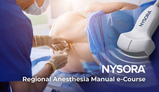
Regional Anesthesia Manual e-Course
(formerly Compendium of RA)
What will you learn?
- Spinal, epidural, and nerve block procedures and management protocols
- How to perform over 60 nerve blocks, with step-by-step instructions on anatomy, indications, technique, choice of local anesthetic, and more.
- The ultimate tips by key opinion leaders on how to succeed every single time.
- The practical side of regional anesthesia, and how to instantly introduce new blocks to your practice.
“As an author of several bestselling books in anesthesiology, I have always been frustrated with by the limitations of the format of books and ebooks. Learning regional anesthesia and ultrasound relies on visuals, animations, and videos which the book/ebook format doesn’t allow.
Besides, by the time a book is published – it’s already outdated and can’t be updated with new information.” – says Dr. Admir Hadzic.
This is why we created the NYSORA Learning System and built everything you need to know about RA in the Regional Anesthesia Manual e-Course.
In the Regional Anesthesia Manual e-Course, the information is enriched with as many visuals, animations, and videos as necessary for faster comprehension and knowledge retention. Plus, we added a note-taking tool so that you can keep all additional information in one place, and a community for information-sharing so that it never feels like you’re learning alone.
Experience the Regional Anesthesia Manual e-Course on the NYSORA LMS, and you’ll never go back to your old books.
- Access on any device
- Real-time updates
- All regional anesthesia procedures and management protocols
- Step-by-step technique instructions
- NYSORA’s fabled functional regional anatomy illustrations and animations
- Reverse ultrasound anatomy videos used in lectures worldwide
- Teaching collections of clinical videos
- Collection of Infographics for exam preparation (e.g. EDRA)
- A mobile app
Course Content
Foundations
Course Includes
- 72 Lessons
- 743 Topics
- 14 Quizzes